Precision Cardiovascular Monitoring with Non-Invasive Hemodynamic Assessment

Introduction

Accurate, real-time assessment of hemodynamic parameters such as blood flow and tissue perfusion is critical for diagnosing and managing cardiovascular diseases. Traditional methods for measuring these parameters, such as catheterization or imaging techniques, can be invasive, time-consuming, and uncomfortable for patients. Our patented technology provides a groundbreaking non-invasive solution for assessing hemodynamics using a coherent light source, allowing for real-time, accurate measurements of blood flow and tissue health without the need for invasive procedures. This innovation stands to transform how healthcare providers monitor and diagnose cardiovascular health.

The Need for Non-Invasive Cardiovascular Monitoring

Cardiovascular diseases are the leading cause of death globally, making early detection and monitoring of vital hemodynamic parameters essential in managing patient outcomes. Traditional diagnostic methods, such as angiography, ultrasound, and invasive pressure monitoring, often come with significant discomfort, risks, and limitations in accessibility. These methods can also be impractical for continuous monitoring, which is needed in critical care settings or for patients with chronic conditions. The healthcare industry requires a less invasive, more accessible way to monitor cardiovascular health in real-time.

For patients, non-invasive monitoring systems would offer a more comfortable, accessible, and continuous method of tracking their cardiovascular health, improving patient adherence and quality of care.

A Breakthrough in Non-Invasive Hemodynamic Assessment

Our patented technology uses a coherent light source to interrogate biological tissue, enabling non-invasive assessment of hemodynamic parameters. By detecting changes in light patterns as they pass through tissues, the system provides real-time data on blood flow, tissue perfusion, and vascular health. This makes it ideal for a range of applications, from monitoring patients in critical care to offering continuous assessments for those with chronic cardiovascular conditions.

The device’s non-invasive nature means it can be deployed in both clinical and home settings, enhancing telemedicine capabilities and enabling remote patient monitoring. With its real-time feedback, this technology is particularly useful for early detection of issues such as ischemia or vascular dysfunction, helping healthcare providers make timely and informed decisions.

Key Benefits

  • Non-Invasive Monitoring: Eliminates the need for invasive procedures, offering a more patient-friendly diagnostic experience.
  • Real-Time Data: Provides immediate insights into hemodynamic parameters, allowing for quicker clinical decisions.
  • Versatile Applications: Useful for both in-hospital and remote monitoring, expanding its utility in telemedicine and chronic care management.
  • Enhanced Cardiovascular Care: Improves the ability to monitor and manage cardiovascular diseases more effectively.

A New Standard for Cardiovascular Diagnostics

Licensing this technology offers medical device manufacturers and healthcare providers an innovative tool for non-invasive, real-time cardiovascular monitoring. With the potential to improve patient outcomes and streamline diagnostics, this technology is poised to become a key asset in modern cardiovascular care.
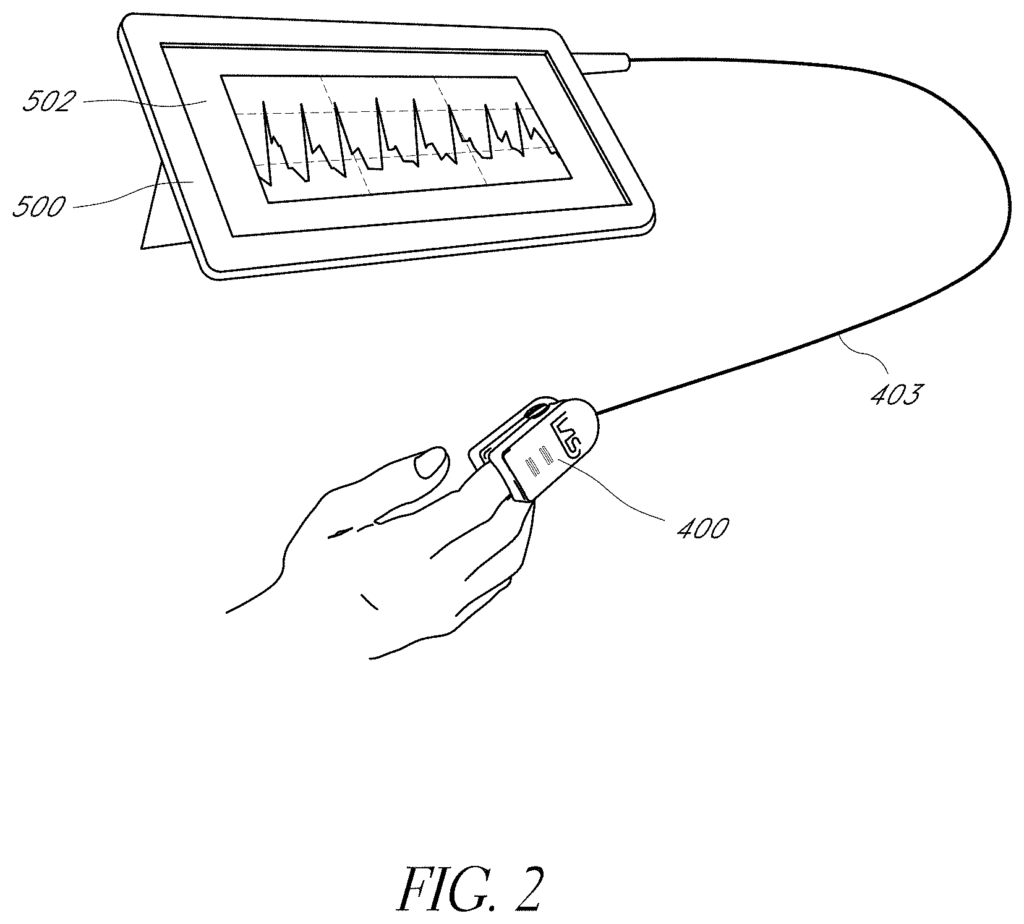
Systems and methods are disclosed for determining physiological information in a subject. The system includes: a light source positionable along a first location outside of the subject; a photo-sensitive detector positionable along a second location outside of the subject and configured to detect scattered light and generate a signal; a processor having a program and a memory, wherein the processor is operably coupled to the detector and configured to receive and store the signals generated over a period of time; wherein the processor is programmed to derive contrast metrics from the stored signals, calculate a waveform from the contrast metrics, decompose the waveform into basis functions and respective amplitudes, and compare the basis function amplitudes to determine the physiological information.

What is claimed is:

1. A system for determining one or more physiological parameters in a subject, the system comprising:

a. a light source positionable along a first location outside of the subject, and configured to direct light from the first location toward a plurality of particles flowing in pulsatile motion within a blood vessel inside of the subject;
b. a photo-sensitive detector positionable along a second location outside of the subject, and configured to detect light from the plurality of particles and generate only one raw signal in response to the detected light, wherein the detected light includes the light scattered and absorbed by the plurality of particles; and

c. a processor comprising a program and a memory, wherein the processor is operably coupled to the photo-sensitive detector and configured to receive and store in memory the raw signal from the photo-sensitive detector generated over a period of time, and wherein the processor is further configured to generate a first waveform and a second, physiologically distinct waveform from the first waveform based on the raw signal;

wherein the processor is programmed to:
i. derive contrast metrics and intensity metrics from the raw signal;

ii. calculate the first waveform from the contrast metrics and the second waveform from the intensity metrics;

wherein the first waveform is a speckleplethysmograph (SPG) waveform and the second waveform is a photoplethysmogram (PPG) waveform;
iii. decompose the first waveform and the second waveform into corresponding one or more characteristic features; and
iv. make a comparison of the corresponding one or more characteristic features of the first waveform and the second waveform to determine the one or more physiological parameters, wherein the one or more physiological parameters are selected from a group consisting of atherosclerotic obstruction, vascular compliance, blood pressure, cardiac output, venous state, vascular tone, blood flow, hemodynamics, and combinations thereof.
2. The system of claim 1, wherein the processor is further programmed to convert the contrast metrics into metrics of volumetric flow and convert the intensity metrics into metrics of volumetric expansion.
3. The system of claim 1, wherein the one or more characteristic features are amplitudes of a basis function and wherein the processor is further programmed to generate a histogram based on a ratio of basis function amplitudes.
4. The system of claim 1, wherein the one or more characteristic features are amplitudes of a periodic basis function, and the decomposition is equivalent to a time-frequency transform.
5. The system of claim 1, wherein the one or more characteristic features are amplitudes of a wavelet basis function, and the decomposition represent a wavelet transform.
6. The system of claim 1, wherein the one or more characteristic features describe widths of the first waveform and the second waveform.
7. The system of claim 1, wherein the one or more characteristic features are timing occurrences of local extrema of the first waveform and the second waveform, and wherein the processor is further programmed to calculate a time delay between the first waveform and the second waveform based on the timing occurrences of local extrema of the first waveform and the second waveform and further determine the one or more physiological conditions based on the time delay.
8. The system of claim 1, wherein the one or more characteristic features are amplitudes of local extrema of the first waveform and the second waveform, and wherein the processor is further programmed to compare one or more of the amplitudes of the local extrema of the first waveform and the second waveform, a difference in amplitudes of local extrema of the first waveform, and a ratio of amplitudes of local extrema of the first waveform and the second waveform to determine the one or more physiological conditions.
9. The system of claim 1, wherein the one or more characteristic features are magnitudes of sloped of the first waveform and the second waveform, and wherein the processor is further programmed to compare magnitudes of slopes of the first waveform and the second waveform to determine the one or more physiological conditions.

10. A method for determining one or more physiological parameters from particles in pulsatile motion within a physiological system, the method comprising:

a. positioning a light source at a first site outside of the physiological system;
b. actuating the light source, such that light is directed toward the particles;
c. positioning a photo-sensitive detector at a second site outside of the physiological system, wherein the second site is located along a path of light scattered and absorbed by at least some of the particles;
d. using the photo-sensitive detector to detect light scattered and absorbed by at least some of the particles over a period of time in response to the directed light and generate only one raw signal based on the detected light;
e. communicating the raw signal related to the detected light to a processor;
f. calculating a contrast metric from the raw signal and an absorption metric from the raw signals;
g. producing a first, contrast waveform based on changed in the contrast metric over time due to the pulsatile motion of the particles within the physiological system, wherein the first, contrast waveform is a SPG waveform;
h. producing a second, absorption waveform based on changes in the absorption metric over the time due to pulsatile expansion of the particles within the physiological system, wherein the second, absorption waveform is a PPG waveform;
i. decomposing the first, contrast waveform and the second, absorption waveform into respective characteristic features;
j. making a comparison of the respective characteristic features decomposed from each of the first, contrast waveform and the second, absorption waveform; and
k. determining the one or more physiological parameters based at least in part on the comparison, wherein the one or more physiological parameters are selected from a group consisting of atherosclerotic obstruction, vascular compliance, blood pressure, cardiac output, venous state, and vascular tone.
11. The method of claim 10, wherein the characteristic features comprise one or more of amplitudes of basis functions.
12. The method of claim 11, wherein decomposing comprises generating a histogram based on a ratio of the amplitudes of the basis functions.
13. The method of claim 11, wherein the characteristic features comprise widths of the first waveform and the second waveform.
14. The method of claim 11, wherein the characteristic features comprise magnitude of slopes of first waveform and the second waveform.
15. The method of claim 11, further comprising determining local extrema of the first waveform and the second waveform by determining time points in the first waveform and the second waveform which experience relative maximum or minimum value over a time period.
16. The method of claim 15, wherein the characteristic features comprise timing occurrences of the local extrema of the first waveform and the second waveform and wherein comparing comprises determining a temporal offset between the first waveform and the second waveform by determining a difference in the timing occurrences of the local extrema of the first waveform and the second waveform.
17. The method of claim 16, further comprising calculating differences in the timing occurrences of the local extrema of the first waveform and the second waveform to determine delays between corresponding local extrema of the first waveform and the second waveform.
18. The method of claim 17, comprising finding peaks in the first waveform and the second waveform using peak-finding algorithms, and further comprising calculating delays between corresponding peaks of the first waveform and the second waveform.
19. The method of claim 18, wherein the one or more physiological parameters comprises vascular tone, and determining the vascular tone comprises determining a degree of constriction of a blood vessel based on the delays.
20. The method of claim 10, wherein the basis functions are periodic, and wherein decomposing further comprises performing a time frequency transform of the amplitudes of the periodic basis functions.
21. The method of claim 10, wherein the characteristic features comprise amplitudes of a wavelet basis function.
22. The method of claim 21, wherein the decomposing further comprises performing a wavelet transform on the wavelet transform function.

Share

Title

Non-invasive hemodynamic assessment via interrogation of biological tissue using a coherent light source

Inventor(s)

Tyler Bywaters Rice, Michael Ghijsen, Bruce J. Tromberg, Bruce Yee Yang, Sean Michael White

Assignee(s)

University of California, Covidien LP

Patent #

10813597

Patent Date

October 27, 2020

Inquire about this intellectual property

Learn more about "Precision Cardiovascular Monitoring with Non-Invasive Hemodynamic Assessment"