Advanced Raman Imaging for Early Caries Detection
Introduction
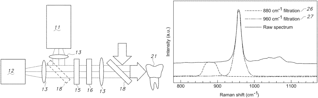
This full-scale Raman imaging technology offers a powerful tool for detecting early-stage dental caries before visible symptoms arise. By using Raman spectroscopy, the system provides precise, non-invasive imaging of tooth structure, identifying areas of decay at the molecular level. For dental professionals and healthcare providers, this technology represents a breakthrough in preventive dentistry, enabling early diagnosis and treatment of cavities, reducing the need for more invasive procedures down the line, and improving overall patient care.
The Challenge: Detecting Dental Decay at an Early Stage
Dental caries (cavities) are one of the most common oral health problems worldwide, often going undetected until significant damage has been done to the tooth. Traditional methods of diagnosing caries, such as visual inspections and X-rays, may not detect early decay, leaving patients vulnerable to more extensive dental procedures. As patient demand grows for less invasive, more accurate diagnostic tools, dental care providers need advanced technology that offers earlier detection and more precise insights into tooth health.
Raman Imaging for Accurate, Non-Invasive Diagnosis
This Raman imaging technology solves these issues by using advanced spectroscopy to detect early-stage caries with high precision. The imaging system provides detailed, real-time images of tooth composition, allowing dental professionals to identify decay before it reaches the advanced stages that require fillings or root canals. The non-invasive nature of the technology makes it ideal for routine dental exams, offering patients a more comfortable and proactive approach to maintaining oral health. Early detection enables timely intervention, which can prevent the progression of decay and preserve the natural structure of the tooth.
Key Benefits for Dental and Healthcare Providers
For dental offices and clinics, this advanced Raman imaging technology offers a competitive advantage by enabling earlier, more accurate caries detection. It enhances the level of care provided to patients, ensuring that cavities are identified and treated in their earliest stages. Dental equipment manufacturers and diagnostic tool providers can integrate this technology into their product lines, offering cutting-edge solutions that align with the increasing demand for non-invasive, high-precision diagnostics in oral healthcare.
Investing in Cutting-Edge Dental Diagnostics
Licensing this advanced Raman imaging technology for caries detection allows your company to lead the way in dental innovation. By offering a system that improves early diagnosis, reduces invasive treatments, and enhances patient care, you position your business at the forefront of preventive dentistry. This technology represents a valuable investment for companies committed to improving oral health outcomes and delivering state-of-the-art diagnostic solutions.
- Abstract
- Claims
I claim:
1. An improved Raman imaging system for obtaining images from a sample surface in seconds comprising:
9. An improved method of obtaining Raman images of a sample surface comprising:
Share
Title
Full scale Raman imaging for early caries detection
Inventor(s)
Shan Yang
Assignee(s)
Jackson State University
Patent #
10876972
Patent Date
December 29, 2020