Advanced Monitoring for Pulmonary Artery Pressure in Critical Care

Introduction

Chronic heart failure and pulmonary artery hypertension are conditions that require meticulous monitoring to prevent sudden complications. Monitoring pulmonary artery pressure (PAP) provides crucial insights into a patient’s cardiovascular and pulmonary status, helping clinicians detect early signs of deterioration and adjust treatment plans accordingly. Our patented pulmonary artery pressure change monitor offers a cutting-edge solution to this ongoing need, providing real-time monitoring that enables early intervention and improved patient outcomes.

Current Limitations in Monitoring Pulmonary Artery Pressure

Traditional methods for monitoring pulmonary artery pressure often involve invasive procedures or intermittent assessments, which limit their usefulness in real-time management of conditions such as heart failure or pulmonary hypertension. These approaches may not provide the continuous data necessary to detect subtle changes in a patient’s condition, leading to delayed interventions and potentially worsening outcomes. For patients who are prone to sudden pulmonary or cardiac complications, real-time monitoring is critical for preventing acute events like heart failure exacerbation or pulmonary edema.

The healthcare industry is moving toward more personalized and real-time approaches to care, where continuous monitoring can provide early warnings and allow for immediate medical adjustments, thereby reducing hospital readmissions and improving quality of life.

A Game-Changing Approach to Pressure Monitoring

Our pulmonary artery pressure change monitor is designed to provide real-time, non-invasive or minimally invasive monitoring of pulmonary artery pressure changes, making it easier to track a patient’s condition over time. By detecting subtle fluctuations in pressure, this system enables healthcare providers to proactively adjust treatments, reduce the risk of complications, and offer more personalized care. The monitor is ideal for use in both hospital settings and at home for remote patient monitoring, making it versatile for a wide range of clinical applications.

This advanced system allows physicians to have continuous insight into a patient’s cardiovascular status, offering real-time data that can inform treatment decisions and reduce the need for more invasive procedures. By integrating this technology into routine care, healthcare providers can offer better management of chronic conditions, improve patient outcomes, and reduce costs related to emergency interventions.

Key Benefits

  • Real-Time Monitoring: Provides continuous data on pulmonary artery pressure, allowing for immediate intervention when necessary.
  • Non-Invasive or Minimally Invasive: Reduces the need for traditional invasive monitoring methods.
  • Improved Patient Management: Enables personalized care plans for chronic conditions like heart failure and pulmonary hypertension.
  • Versatile Use: Suitable for both in-hospital care and remote monitoring at home, expanding its applicability in modern healthcare.

Advancing Cardiovascular Care with Real-Time Insights

Licensing this pulmonary artery pressure monitor technology offers medical device companies and healthcare providers an essential tool for advancing patient care in cardiology and pulmonology. With its ability to provide continuous, real-time data, this technology is critical for improving patient management and outcomes in chronic cardiovascular and pulmonary conditions.
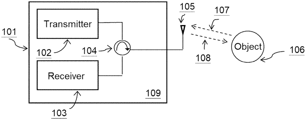
The present disclosure describes systems, methods, and devices to infer changes in pulmonary artery pressure in a subject using Doppler radar. A portable, non-invasive device for non-invasively measuring right ventricular cardiac motion that can be used in a subject’s home can infer pulmonary artery pressure changes to increase patient compliance and mitigate the likelihood of heart decompensation. A mobile pulmonary artery pressure monitor can be especially useful to patients with congestive heart failure who are elderly, incapacitated, or do not have easy access to a clinic, doctor’s office, or hospital.

What is claimed is:

1. A method of detecting a heart failure condition in a heart in a body of a subject, comprising:

a) obtaining a first average motion depth of a right ventricle of the heart over a first time period of detection, wherein the first average motion depth is based on an analysis of a first electromagnetic signal that enters the body of the subject from outside the body, is reflected off the right ventricle of the heart, exits the body, and is then detected outside the body;
b) obtaining a second average motion depth of the right ventricle of the heart over a second time period of detection, wherein the second average motion depth is based on an analysis of a second electromagnetic signal that enters the body of the subject from outside the body, is reflected off the right ventricle of the heart, exits the body, and is then detected outside the body;
c) determining a change between the first average motion depth of the first motion average depth of the right ventricle of the heart over the first time period of detection and the second average motion depth of the second average motion depth of the right ventricle of the heart over the second time period of detection; and
d) determining, based on the change between the first average motion depth of the first motion average depth of the right ventricle of the heart over the first time period of detection and the second average motion depth of the second motion average depth of the right ventricle of the heart over the second time period of detection, whether the subject has the heart failure condition,
wherein the heart failure condition comprises decompensated heart failure,
wherein the first electromagnetic signal and the second electromagnetic signal are radio waves.
2. The method of claim 1, wherein the first time period of detection is a single heartbeat of the subject.
3. The method of claim 1, wherein the second time period of detection is a single heartbeat of the subject.
4. The method of claim 1, wherein the heart failure condition further comprises acute decompensated heart failure.
5. The method of claim 1, wherein the heart failure condition further comprises congestive heart failure.
6. The method of claim 1, wherein the heart failure condition further comprises fluid accumulation in the heart.
7. The method of claim 1, wherein the heart failure condition further comprises right-sided heart failure.
8. The method of claim 1, wherein the heart failure condition further comprises left-sided heart failure.
9. The method of claim 1, wherein the heart failure condition further comprises systolic heart failure.
10. The method of claim 1, wherein the heart failure condition further comprises diastolic heart failure.
11. The method of claim 1, wherein the heart failure condition further comprises Stage A heart failure.
12. The method of claim 1, wherein the heart failure condition further comprises Stage B heart failure.
13. The method of claim 1, wherein the heart failure condition further comprises Stage C heart failure.
14. The method of claim 1, wherein the heart failure condition further comprises Stage D heart failure.
15. The method of claim 1, wherein the subject is human.
16. The method of claim 1, wherein the first average magnitude of the first average motion depth of the right ventricle is a first average amplitude of contraction of the right ventricle, wherein the second average motion depth of the right ventricle is a second average amplitude of contraction of the right ventricle.
17. The method of claim 1, wherein the first average magnitude of the first average motion depth of the right ventricle is a first average amplitude of expansion of the right ventricle, wherein the second average motion depth of the right ventricle is a second average amplitude of expansion of the right ventricle.
18. The method of claim 1, wherein the change between the first average motion depth of the right ventricle of the heart over the first time period of detection and the second average motion depth of the right ventricle of the heart over the second time period of detection is associated with a pulmonary artery pressure that is associated with the heart failure condition.
19. The method of claim 1, wherein the change between the first average depth of the right ventricle of the heart over the first time period of detection and the second average motion depth of the right ventricle of the heart over the second time period of detection is a decrease.
20. The method of claim 19, wherein the decrease between the first average motion depth of the right ventricle of the heart over the first time period of detection and the second average motion depth of the right ventricle of the heart over the second time period of detection indicates an increase in the pulmonary artery pressure in the subject.
21. The method of claim 1, wherein the change between the first average magnitude of the first average motion depth of the right ventricle of the heart over the first time period of detection and the second average motion depth of the right ventricle of the heart over the second time period of detection is an increase.
22. The method of claim 21, wherein the increase between the first average motion average depth of the right ventricle of the heart over the first time period of detection and the second average motion depth of the right ventricle of the heart over the second time period of detection indicates a decrease in the pulmonary artery pressure in the subject.

Share

Title

Pulmonary artery pressure change monitor

Inventor(s)

Xiaomeng Gao, Xiaonan JIANG, Xiaoguang Liu, Dennis Matthews, Saul Schaefer

Assignee(s)

University of California

Patent #

11647978

Patent Date

May 16, 2023

Inquire about this intellectual property

Learn more about "Advanced Monitoring for Pulmonary Artery Pressure in Critical Care"