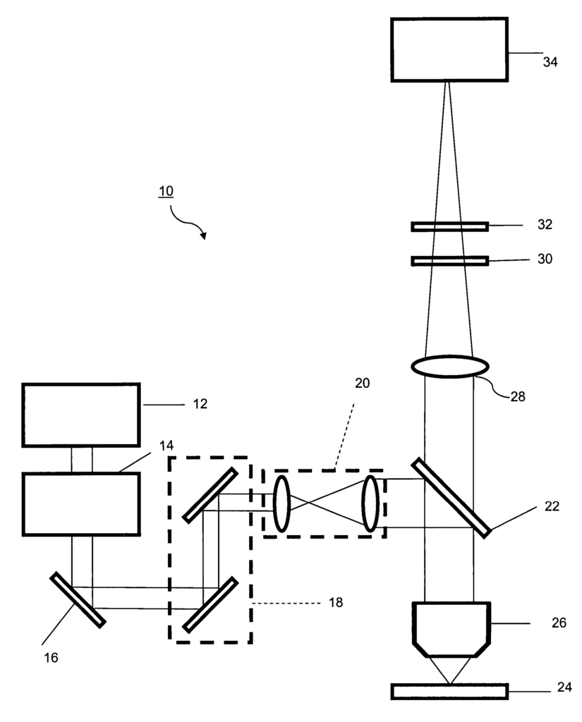
Precision Imaging with Two-Photon Microscope Technology
Introduction
In the fields of biotechnology, medical research, and diagnostics, high-resolution imaging is crucial for understanding complex biological processes and discovering new therapies. However, traditional imaging techniques often fall short when it comes to deep tissue penetration and achieving molecular-level detail without damaging live samples. Our patented two-photon microscope with spectral resolution offers a cutting-edge solution, providing unprecedented imaging depth and clarity that can transform the way researchers visualize biological structures in live tissue.
Current Gaps in Imaging Technology
Conventional microscopes, while useful for many research purposes, struggle with limitations in penetration depth and resolution when applied to thick or live tissue samples. Additionally, imaging live tissues can result in sample damage due to high-intensity light exposure. This is particularly problematic in fields like neuroscience and cancer research, where precise, non-invasive imaging is essential for observing biological processes in real time. Furthermore, standard imaging systems lack spectral resolution, making it difficult to distinguish between different molecular signatures within a sample.
These limitations create barriers for researchers who require more detailed and precise imaging technologies to gain meaningful insights from their work. The need for a high-performance system capable of imaging deep into live tissues, while maintaining both clarity and spectral resolution, is clear across many scientific disciplines.
A Breakthrough in Spectral Imaging with Two-Photon Microscopy
Our patented two-photon microscope with spectral resolution addresses these challenges by enabling high-resolution, deep tissue imaging while minimizing sample damage. Two-photon excitation allows for deeper penetration into tissues with less photodamage, making it ideal for imaging live cells, tissues, and even entire organisms. The addition of spectral resolution further enhances the technology by allowing researchers to separate and analyze distinct molecular signals, providing a more detailed view of complex biological structures.
This innovative system can be used in a wide range of applications, from mapping neural networks in the brain to studying the behavior of cancer cells. Its non-invasive nature makes it a powerful tool for live imaging in real-time, offering researchers greater flexibility and accuracy in their studies. Additionally, the spectral capabilities enable the differentiation of various fluorophores or molecular markers, unlocking new possibilities in multi-modal imaging.
Key Advantages
- Deep Tissue Penetration: Two-photon excitation allows for clear imaging of live tissues without damaging the sample.
- Spectral Resolution: This technology can separate molecular signals, enabling the study of multiple markers in a single sample.
- Non-Invasive Imaging: The system is ideal for live imaging, reducing photodamage and allowing for real-time observations.
- Wide Range of Applications: From neuroscience to cancer research, this microscope opens up new possibilities for advanced research.
A Leap Forward in Imaging Precision
Licensing this two-photon microscope technology with spectral resolution provides research institutions, biotechnology companies, and pharmaceutical developers with a highly advanced imaging tool. With applications spanning from basic research to drug discovery, this technology enables scientists to visualize biological processes in unprecedented detail, offering a significant edge in the race to make new breakthroughs.

- Abstract
- Claims
1. A microscope for generating a multi-dimensional, spectrally resolved image of a dynamic sample, the microscope comprising:
11. A microscope for generating an image of a sample, the microscope comprising:
12. A method of generating an image of a sample having x and y dimensions and using a microscope having a laser light source, a computer controlled scanning mirror, a dispersive element and a camera, the method comprising:
17. A method of generating an image of a sample having x and y dimensions and using a microscope having a laser light source, a computer controlled scanning mirror, a dispersive element and a camera, the method comprising:
Share
Title
Two-photon microscope with spectral resolution
Inventor(s)
Valerica Raicu, Russell Fung
Assignee(s)
UWM Research Foundation Inc
Patent #
7973927
Patent Date
July 5, 2011
